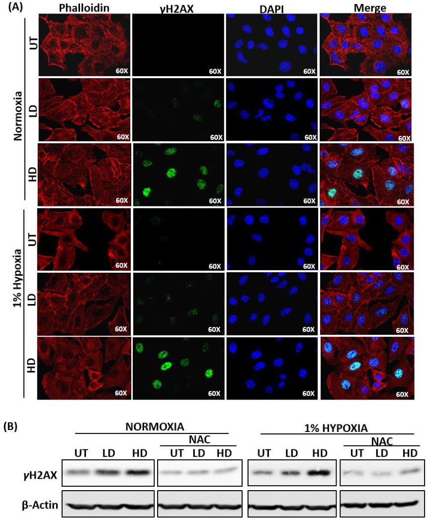
Fig. 4. Accumulation of DNA damage in Allicin treated A549 cells. (A) Confocal images of A549 cells expressing γ-H2AX after treatment with 10μg/ml (LD) and 40μg/ml (HD) allicin for 24hr in normoxic and hypoxic condition at 60X magnification. Phalloidin was used to stain actin fibres and DAPI was used to stain nuclei (B) Western blot analysis of γ-H2AX in A549 cells treated with 10μg/ml (LD) and 40μg/ml (HD) allicin in absence and presence of antioxidant NAC and cultured in normoxia and 1% hypoxia for 48hr. UT= untreated.